


Doctors from Boston Children’s Hospital and Brigham and Women’s Hospital have performed the first embolization of a vein of Galen malformation on an unborn infant, according to research published Thursday in the Stroke journal.
According to Boston Children’s “a vein of Galen malformation (VOGM) is a type of rare blood vessel abnormality inside the brain.”
Those with VOGM have misshapen arteries in the brain that connect directly with veins instead of with capillaries, the hospital explained. Since capillaries help slow blood flow, the malformation “causes a rush of high-pressure blood into the veins.”
It poses several risks for infants, including congestive heart failure, pulmonary hypertension, brain injury and hydrocephalus.
“Despite decades of technique refinement of trans-arterial embolization and the establishment of specialty referral centers, fetuses diagnosed with vein of Galen malformation continue to have high mortality, with survivors facing high rates of severe neurological and cognitive morbidity,” said the study.
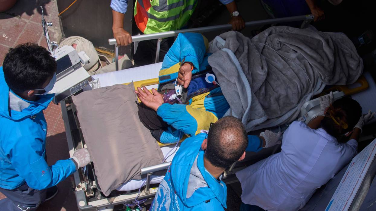
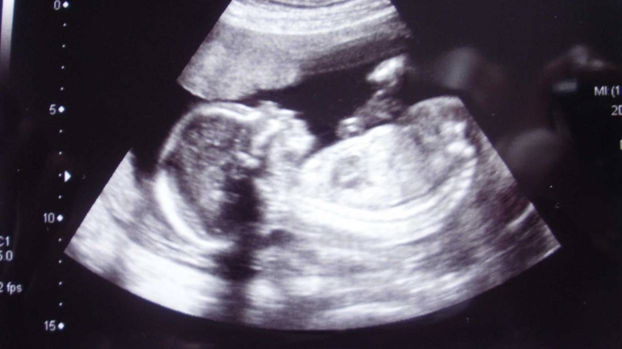
With the assistance of a transuterine ultrasound, researchers performed an in-utero embolization – a procedure described by the Cleveland Clinic as “placing a substance in [a] vessel to prevent blood from flowing through it” – to address VOGM in a fetus 34 weeks and two days gestational age. It was the first treated patient in a clinical trial that is underway at the Boston hospitals and it was performed with oversight from the U.S. Food and Drug Administration.
“Fetal ultrasound showed an immediate drop in abnormal blood flow through the brain malformation, and fetal echocardiography showed significant improvement in heart function the day after the procedure,” said the American Heart Association.
There was a premature rupture of the mother’s membranes during the procedure and the infant was delivered two days later. Her newborn baby did not require any cardiovascular support or surgery following the in-utero treatment. They were watched in the NICU for several weeks due the premature birth and sent home.
Since it was born, the infant has required no medication to treat heart failure and no postnatal surgery to treat the malformation. Echocardiograms have shown improvement in cardiac output, brain MRIs showed no brain injury and neurological exam was normal.
“In our ongoing clinical trial, we are using ultrasound-guided transuterine embolization to address the vein of Galen malformation before birth, and in our first treated case, we were thrilled to see that the aggressive decline usually seen after birth simply did not appear. We are pleased to report that at six weeks, the infant is progressing remarkably well, on no medications, eating normally, gaining weight and is back home. There are no signs of any negative effects on the brain,” said lead study author Darren B. Orbach, co-director of the Cerebrovascular Surgery & Interventions Center at Boston Children’s Hospital and an associate professor of radiology at Harvard Medical School.
Estimates indicate that VOGM is “the most common congenital vascular brain malformation,” and that it occurs in as many as one in every 60,000 births, per the AHA. Current standard treatments are performed after birth.
“This approach represents a paradigm shift in management of this challenging condition,” said the research of the new in-utero approach.
“The key advance here is to intervene before the physiologic events of birth can cause life-threatening heart failure,” said Colin P. Derdeyn, a neurointerventional radiologist at University of Iowa Health Care who performs VOGM embolizations on neonates. He was not involved with the study. “There are caveats; one successful case is not enough experience for us to conclude that the risks of this procedure are worth the benefits. Safety issues may crop up in future procedures, and this approach through the veins may not be consistently successful in preventing heart failure. The procedure described here is designed to reduce the flow through the malformation and not to cure it.”
More procedures of its kind will need to be performed as part of the clinical trial, explained Gary M. Satou, the director of pediatric echocardiography at UCLA Mattel Children's Hospital and co-director of the UCLA Fetal Cardiology Program. He was also not involved with the study.
“The national clinical trial will be crucial in order to achieve adequate data and, hopefully, successful outcomes,” he said.