A map of a fruit fly’s brain released this week could help scientists researching cures and treatments for Alzheimer’s disease and Parkinson’s disease, according to the Nature journal.
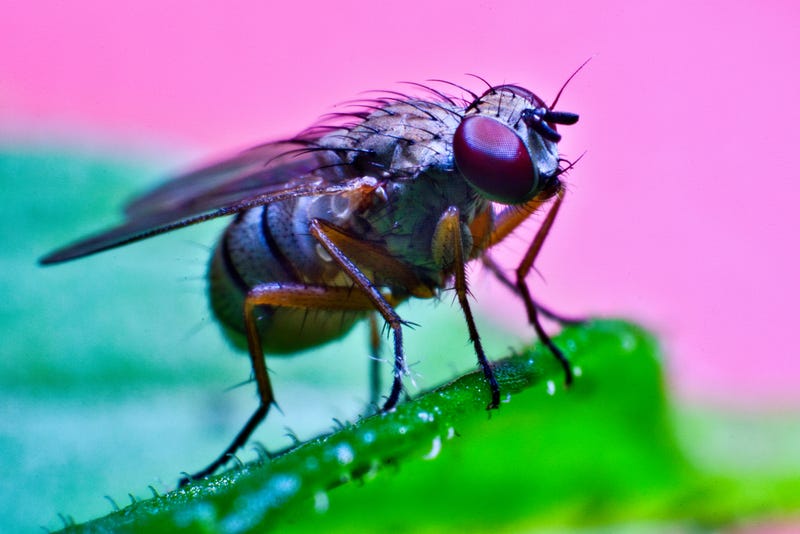
“Scientists have generated the first complete map of the brain of a small insect, including all of its neurons and connecting synapses,” said an article in the journal. That inspect was the fruit fly Drosophila melanogaster, which has a brain smaller than a poppy seed.
Even so, researchers were able to map all 3,016 neurons and 548,000 synapses “tightly packed” in the insect’s brain. This is a “milestone in understanding how the brain processes the flow of sensory information and translates it into action,” Nature explained.
Now that scientists have this model to use as a reference brain, they can “look at what happens to connectivity in models of Alzheimer’s and Parkinson’s diseases and of any degenerative disease,” said Marta Zlatic, a neuroscientist at the University of Cambridge, U.K., and co-author of the research paper.
Alzheimer’s disease is a form of dementia. It is linked to plaques – protein deposits called beta-amyloid that build up in the space between nerve cells – and tangles – twisted fibers of a protein called tau that build up inside cells – according to the Alzheimer’s Association.
Parkinson’s disease “is a brain disorder that causes unintended or uncontrollable movements, such as shaking, stiffness, and difficulty with balance and coordination,” according to the National Institute on Aging.
In patients who have Parkinson’s disease, nerve cells in the basal ganglia (an area of the brain that controls movement) that produce dopamine become impaired or die. Scientists do not know what causes the neurons to die, said the institute. However, many people with the condition have Lewy bodies, which are unusual clumps of the protein alpha-synuclein.
To create the fruit fly brain map, researchers spent a year and a half capturing images of the brain of a single six-hour-old larva with a nanometre-resolution electron microscope. Next, they used a computer-assisted program to pinpoint synapse, followed by a manual process of double-checking.
Kei Ito, a neuroscientist at the University of Cologne, Germany, explained that the process was “really time-consuming and labor-intensive.”
Now that one brain has been mapped, it should be easier to map more in the future, including brains of other species.
“One can now use it to train machine learning to do it much faster,” said Zlatic. Next, researchers are looking to map the brain of an adult Drosophila melanogaster.


